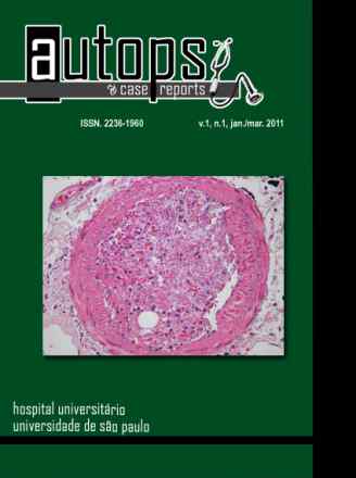
Lung adenocarcinomas with a mixture of tubular or papillary pattern, sheet-like or trabecular architecture, eosinophilic cytoplasm with centrally located nuclei and alpha-fetoprotein-producing cells have been described as hepatoid adenocarcinomas. Hepatoid adenocarcinomas are mainly found in the stomach but rare cases in other organs have been described. Immunostaining for alpha-fetoprotein (AFP), hepatocyte paraffin 1 (HepPar-1) and thyroid transcription factor-1 (TTF-1) helps in the diagnostic workup. Tumor behavior is still not entirely known but it seems to be associated with early metastases. We report on a 66-year-old, heavy-smoker male patient who had a 10-month history of respiratory complaints and weight loss. At the time he was hospitalized, respiratory failure was already established. The computed tomography corresponded to a collapsed right lung due to a poorly defined expanding mass. The bronchoscopy revealed narrowing of the inferior and medium lobar bronchi. The patient developed irreversible shock and died. At the right lung inferior lobe was extensively replaced by a grayish diffuse neoplasia in a “pneumonia-like†gross pattern. Metastatic disease was found in
the right adrenal gland and thoracic and abdominal lymph nodes. Microscopic dissemination through lymphatics, pleura, and airways was detected. Histological examination revealed a poorly differentiated adenocarcinoma with hepatoid features. Immunohistochemmistry stains were positive for keratin 7, polyclonal carcinoembryonic antigen (CEA) in a diffuse pattern, AFP and HepPar-1 antibody. TTF-1 showed a diffuse granular cytoplasmic staining of the neoplastic cells, and only focal nuclear positivity. Multiple bilateral emboli originated from deep venous thrombosis were present in large and medium
branches of the pulmonary artery and contributed to the cause of death.